2347的問題,透過圖書和論文來找解法和答案更準確安心。 我們找到下列免費下載的地點或者是各式教學
2347的問題,我們搜遍了碩博士論文和台灣出版的書籍,推薦弗雷德.希爾寫的 當代日本海上力量 和周伯翰的 生物技術專利要件與商業方法專利適格性之研究都 可以從中找到所需的評價。
這兩本書分別來自風格司藝術創作坊 和元照出版所出版 。
國立陽明交通大學 機械工程系所 王啟川所指導 莫尼實的 超疏水性在結露狀況下對氣冷式熱交換器性能的影響 (2021),提出2347關鍵因素是什麼,來自於熱交換器、超疏水性鰭片、凝結水脫落、熱傳、節能。
而第二篇論文高雄醫學大學 醫藥暨應用化學系博士班 王志光 教授所指導 Swathi Nedunchezian的 運用仿生支架進行骨軟骨修復組織工程的生物設計策略 (2021),提出因為有 透明質酸、明膠、混合水凝膠、3D 生物陶瓷腳手架、軟骨組織工程的重點而找出了 2347的解答。
當代日本海上力量
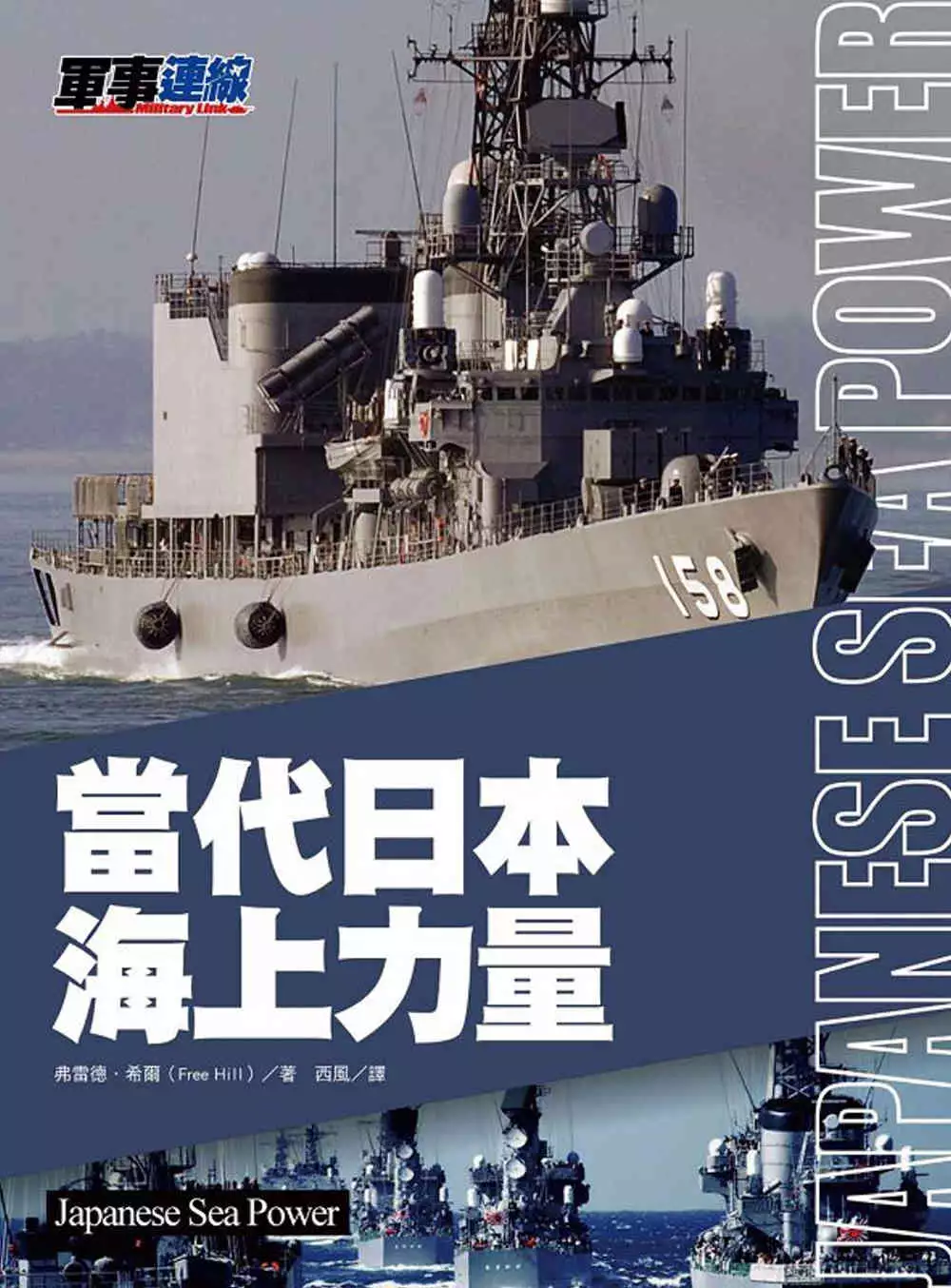
為了解決2347 的問題,作者弗雷德.希爾 這樣論述:
當代日本的海上力量是由日本海上自衛隊組成。日本海上自衛隊,英文為Japan Maritime Self Defense Force,縮寫為JMSDF,是防衛省的下屬特別機關,相當於其他國家的海軍。由於原則上日本不能擁有軍隊,採取防守自衛的立場,因此不能配備大型戰艦、航空母艦以及核動力潛艇,其主要任務是防衛日本領海。 冷戰後日本海上自衛隊不斷發展,除了自衛外,日本自衛隊還能參與到國際救援等國際軍事行動中。目前日本海上力量擁有直升機航空母艦、神盾級驅逐艦等戰鬥力強大的艦船。 本書從日本當代海軍概述講起,描述了日本海軍的組成架構、艦隊組成、作戰指導思想、發展方向、
主要基地等。詳述了大型水面戰艦、水面主力戰艦、潛艇部隊等日本主要海上力量,《當代日本海上力量》還詳細列出了每種戰艦的作戰性能和技術參數,附有精美插圖。
2347進入發燒排行的影片
今日もみてくれてありがとう!!
今回は、pretty noobでゲスト出演したフレッシュさんの撮影会の様子をお届け!
グラビアアイドルの皆さんの水着のポイントを大調査(^○^)/
みんなが気になる撮影会の裏側を教えちゃうよ♡
▽Twitter
https://twitter.com/dpandaramu?s=20
▽Instagram
→https://www.instagram.com/dpandaramu/
▽Tik Tok
→https://www.tiktok.com/@ramu.tiktok?lang=ja-JP
▽Youtube
https://www.youtube.com/channel/UCEdiMA-7EUoUzQAXAJDRZUg/featured
#RaMu
#グラビアアイドル
#水着
#撮影会
#プール
超疏水性在結露狀況下對氣冷式熱交換器性能的影響
為了解決2347 的問題,作者莫尼實 這樣論述:
濕空氣冷凝是熱管理系統中常見的過程,在冷凍空調循環中尤為重要,冷凝現象發生於當熱交換器,特別是蒸發器,在低於空氣露點的溫度下操作時。此現象將會導致鰭片側的冷凝液滴(膜)滯留(retention)與橋接(bridging),進而造成風機壓降與能耗的增加。本研究旨在開發一種超疏水熱交換器,通過其疏水特性,最大限度地減少冷凝水的滯留和橋接。本研究提出一種新型的超疏水性鰭片換熱器設計構想,採用傾斜鰭片排列以達到最小壓降和最大節能效果。本研究從熱傳與壓降性能的觀點切入,將新型超疏水性傾斜鰭片換熱器與其他換熱器作比較分析,分別為:超疏水水平鰭片換熱器、親水性傾斜鰭片換熱器、與親水性水平鰭片換熱器。此外,
本研究藉由改變不同的操作條件,如:進氣溫度、相對濕度和鰭片間距,對這四種換熱器進行性能測試。親水和超疏水換熱器中分別以膜狀冷凝和滴狀冷凝模式為主。由於其表面的高潤濕性,親水換熱器會有較大的液滴脫落直徑。相比之下,超疏水換熱器中發生的 Cassie-Baxter 液滴模式,促使了較小的液滴脫落直徑。本研究建立了一個力平衡模型來分析液滴脫落直徑,模型參數包括了表面張力、慣性力與重力對液滴的影響。本研究基於韋伯數(We)與邦德數(Bo)與液滴脫落直徑,引入了一個新的無因次參數( ),該無因次參數 可預測表面的凝結水脫落能力,在給定的鰭片間距下, 越小代表凝結水脫落能力越好。研究結果表明,滴狀冷凝的
超疏水換熱器在濕空氣下的冷凝熱傳性能相較膜狀冷凝的親水性換熱器並未有顯著的提升,此結果可歸因於非凝結性氣體效應。然而,在壓降方面,超疏水性換熱器與親水性換熱器相比,可帶來高達70%的壓降降低,大幅提升節能效果。壓降的降低歸因於聚結誘發的液滴跳躍現象,使得冷凝水連續脫落。
生物技術專利要件與商業方法專利適格性之研究
為了解決2347 的問題,作者周伯翰 這樣論述:
本書係對於美國專利商標局公布的「基於最高法院在Alice案之決定的初步檢驗說明」與該局在2018年修正公布的「專利審查程序手冊」中,有關如何判斷「專利標的之適格性」與如何適用「標的適格性之兩部標準」進行的說明;美國聯邦最高法院與美國聯邦巡迴上訴法院之相關重要案例建立的判斷標準;台灣智慧財產局公布的《專利審查基準》第2篇第2章、第12章及第14章之相關說明;及智慧財產法院之相關重要案例建立的判斷標準,加以分析闡釋,期能使法學界、司法實務界、專利實務界與產業界對於涉及自然法則、自然現象或抽象概念之標的取得專利的相關標準及法制之重點能有所瞭解,從而使社會大眾對於生物技術與商業
方法之利用能更為合理與普及。
運用仿生支架進行骨軟骨修復組織工程的生物設計策略
為了解決2347 的問題,作者Swathi Nedunchezian 這樣論述:
Acknowledgment iii摘要 vAbstract viiList of figures xiii1. Chapter One 1Introduction 11.1 Problem statement 11.1.1 Articular cartilage 31.1.2 Structure and composition of articular cartilage 31.1.3 Articular cartilage defect 51.2. Surgical techniques for cartilage and Osteochondral repair
currently in use 61.2.1 Bone marrow techniques 61.2.2 Mosaiplasty 81.2.3 Autologous chondrocyte implantation method 91.2.4 Matrix induced autologous chondrocyte implantation 111.3. Tissue engineering approaches to Osteochondral defect repair 121.3.1 Scaffold and hydrogel-based cell delivery 1
41.4. Cell source for tissue engineering purposes 161.4.1 Chondrocyte cells 161.4.2 Adult somatic stem cells 171.4.3 Bone marrow-derived stem cell (BMSCs) 181.4.4 Adipose-derived stem cells (ADSCs) 191.5 Scaffolds and hydrogels for tissue engineering 211.5.1 Natural hydrogels in cartilage tiss
ue engineering 251.6. Crosslinking of hydrogel for tissue engineering purpose 291.6.2 Silicon-dioxide Nanoparticle as crosslinkers in tissue engineering 341.6.3 Interaction of SiO2 nanoparticle with adipose-derived stem cells 361.7 Bio ceramics for Osteochondral tissue engineering and regenerati
on 371.7.1 Bio ceramics in Tissue engineering applications 371.7.2 Applications of bioceramics in Osteochondral tissue engineering 391.8 Research Objectives 421.8.1 The specific aims of this thesis are as follows: 43Chapter Two 44Characteristic and chondrogenic differentiation analysis of hybr
id hydrogels comprise of hyaluronic acid methacryloyl (HAMA), gelatin methacryloyl (GelMA), and the acrylate functionalized nano-silica crosslinker 442.1 Introduction 442.2 Materials and methods 522.2.1 Materials 522.2.2 Synthesis of HAMA hydrogel 522.2.4 Synthesis of acrylate functionalized nS
i crosslinker (AFnSi) 532.2.5 Identification of the synthesis HAMA and GelMA 542.2.6 Production of hybrid hydrogels 552.2.7 Identification of the synthesis AFnSi cross-linker 552.2.8 Fabrication of HG hybrid hydrogels 562.2.9.Swelling ratio evaluation 562.2.10 The microstructure morphology ana
lysis 572.2.11 Mechanical properties evaluation 572.2.12 In vitro degradation assay by hyaluronidase 582.2.13 Isolation and culturing of hADSCs 592.2.14 Cell viability assay 602.2.15 Chondrogenic marker gene expression 612.2.15 Quantification of DNA, sGAG deposition and collagen type Ⅱ synthes
is 622.2.16 Statistical analysis 632.3. Results and Discussion 632.3.1.Identification of the synthesis HAMA and GelMA 632.3.2 Identification of the AFnSi crosslinker 672.3.3 Swelling ratio of HG hybrid hydrogels 702.3.4 Morphological examination of HG hybrid hydrogels 722.3.5 Compressive stud
y of HG hybrid hydrogels 752.3.6.Viscoelastic property of HG hybrid hydrogel 782.3.7. Degradation study of HG hybrid hydrogels 812.3.8.Cell viability evaluation of hADSCs on HG hybrid hydrogels 822.3.8. Chondrogenic differentiation ability of HG hybrid hydrogels 852.4. Conclusion 90Chapter Thr
ee 92Multilayer-based scaffold for Osteochondral defect regeneration in the rabbit model 923.1 Introduction 923.2 Materials and methods 963.2.1 Preparation and Characterization of the 3D bioceramic scaffold by DLP method 963.2.2 Cell isolation and culture 973.2.3 Fabrication of the cell-laden
hydrogel/ 3D bioceramic scaffolds mimicking the Osteochondral tissue. 983.2.4 Surgery 983.2.5 Macroscopic Examination 993.2.6 Tissue Processing for paraffin block 993.2.7 Histological and Immunohistochemical Evaluation 1003.2.8 Masson’s trichrome stain 1013.3 Results and discussion 1023.3.1 C
haracterization of the 3D bioceramic scaffold by DLP method 1023.3.2 Fabrication of the hydrogel with hADSCs into the 3D bioceramic scaffold 1043.3.3 In-vivo studies using rabbit as an animal model 1053.3.5 Histological evaluation of neocartilage formation 1073.3.6 Masson’s trichrome staining an
alysis for neocartilage formation 1093.4. Conclusion 110Chapter four 1104.1 General discussion 1124.2 Future work 1134.2.1 Macroscopic Observation of neocartilage formation for 8 weeks 1145.Reference 115